
Ceph X-Ray: What It Shows and What to Ask Before Agreeing
Learn what a ceph X-ray shows, how it differs from panoramic imaging, and what to ask about orthodontic need, radiation, records and fees.
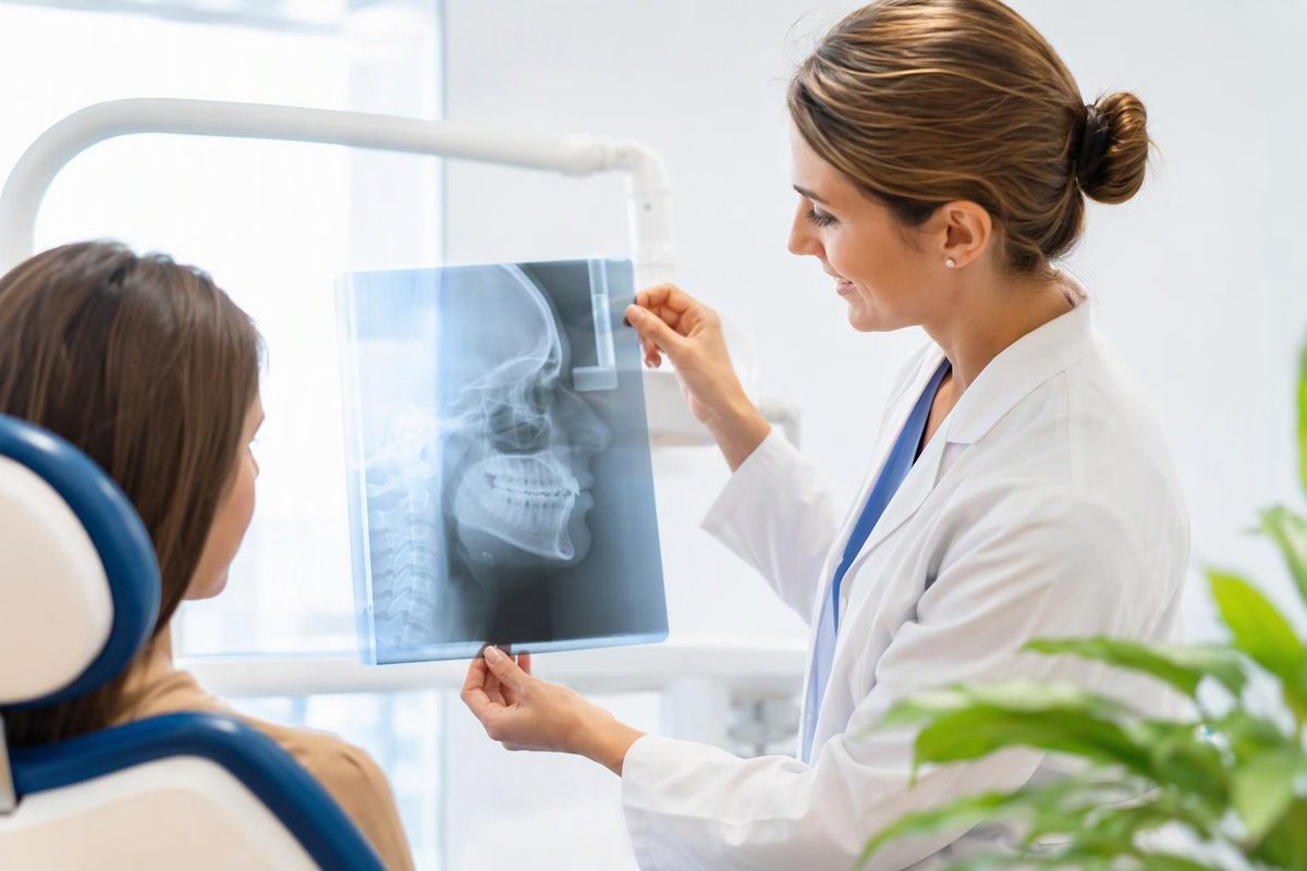
A ceph X-ray, short for cephalometric X-ray, usually means a two-dimensional side-view image of your head. It shows the teeth, jaws and skull together so an orthodontist can assess how they relate—not just whether your teeth look straight. It is commonly used in orthodontic treatment planning. American Association of Orthodontists
If one appears on your braces or aligner quote, the useful question is: “What will this image help you decide in my case?”
Why an orthodontist may recommend it
A side-view ceph can help distinguish a bite problem caused by tooth positions from one involving the relationship between the upper and lower jaws—or both. It can also support planning for corrective jaw surgery. The clinician may trace anatomical landmarks digitally or by hand, then measure angles and distances to help interpret the image. Dental Protection’s guide to lateral cephalometric radiographs
The January 2026 ADA and American Academy of Oral and Maxillofacial Radiology recommendations say a lateral cephalogram may be used to assess the severity of Class II or Class III malocclusion: bite relationships in which the upper and lower teeth or jaws do not line up normally from front to back. It may also help assess associated vertical relationships. These are case-specific indications, not a requirement that every orthodontic patient receive the same images. 2026 imaging recommendations
Ask the clinician to show you what the measurements mean for your treatment options. You do not need to interpret the tracing yourself. If you are still choosing a provider, our guide to dentists versus orthodontists explains their different roles.
Ceph versus panoramic X-ray or CBCT
These tests answer different questions:
| Image | Main view and purpose |
|---|---|
| Lateral ceph | Side profile of the head; helps assess tooth and jaw relationships. |
| Panoramic X-ray | Broad view of the mouth and jaws; useful for assessing tooth eruption and impacted teeth. |
| CBCT | Three-dimensional imaging; may be useful when a complex problem needs information that two-dimensional images cannot provide. |
The AAO’s imaging guide explains these differences. A ceph also does not replace the detailed views used to check for cavities or individual tooth-root problems.
If both a panoramic image and a ceph are proposed, ask what each adds. If CBCT is proposed instead, ask why lower-exposure imaging would not answer the clinical question—the ADA recommends reserving CBCT for situations where those options are insufficient.
What happens during the appointment
The image is taken from outside your mouth, rather than with a sensor placed between your teeth. Staff position your head using supports, tell you how to hold your bite and ask you to stay still. Standardized positioning helps make the image suitable for measurement. Cleveland Clinic’s overview of dental X-rays; positioning and analysis
You may be asked to remove glasses, earrings, hair clips or other objects that could obscure the image. Dental Scan’s patient preparation guidance lists these items. Tell your imaging team beforehand if positioning is difficult, you need accommodations or you are pregnant. Follow your office’s specific preparation instructions.
Radiation and repeat images
Dental X-rays use low levels of radiation, but exposure still needs a clinical justification. The important protection is selecting the right image, using appropriate settings and avoiding unnecessary repeats. For children, the ADA specifically supports individually selected images and child-sized exposure settings. Cleveland Clinic; ADA radiation guidance
Before a second consultation, ask your previous office to send existing images and their dates. The new clinician should review available imaging before deciding whether another exposure is needed; an older or unsuitable image may not answer the current question. 2026 imaging recommendations
Under its 2024 U.S. safety recommendations, the ADA no longer recommends routine lead aprons or thyroid collars for dental imaging, although applicable local rules still govern practice. Their absence alone does not establish unsafe care. ADA shielding guidance
What to check on the bill
Before authorizing orthodontic records, request a written estimate and ask:
- Is the ceph included in the consultation or records fee, or charged separately?
- Does that price include measurement, analysis and an explanation of the findings?
- Are photographs, a panoramic X-ray or a digital tooth scan separate charges?
- Will I owe the records fee if I decide not to start treatment?
- What procedure code will you submit, and what does my insurer say it covers?
For a second opinion, also ask whether the office can use your existing records. Compare the total records charge and what it includes, not just the price beside “ceph X-ray.”