
What to Expect When a Dentist Treats the Pulp but Keeps the Tooth
A 10-step sequence explains how crown pulp is removed, root pulp preserved and the tooth sealed—and why uncontrolled bleeding may redirect treatment.
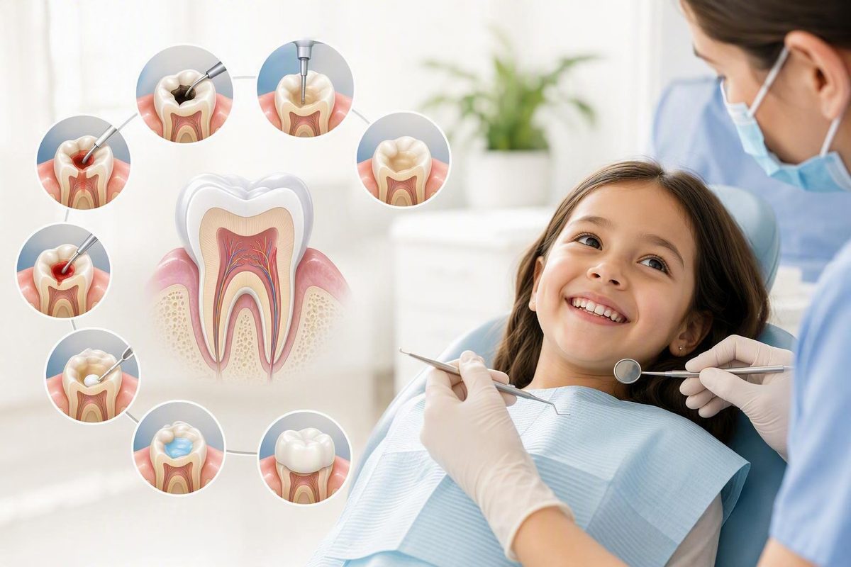
A pulpotomy procedure removes unhealthy or inflamed pulp from the crown of a tooth while attempting to preserve living pulp inside its roots. It is most often discussed when a child has deep decay in a primary molar, although selected permanent teeth can also receive this form of vital pulp therapy.
A pulpotomy is both a treatment and an assessment. The dentist may plan to retain the root pulp, but the condition of the tissue after the tooth is opened—especially whether it appears viable and whether bleeding can be controlled—helps determine whether that plan remains appropriate.
Pain relief, disease control, and tooth retention are goals, not guarantees. Online information cannot establish whether an individual tooth is suitable for treatment. That requires an examination and, commonly, dental radiographs.
Related: How Much Tooth Is Needed for a Crown? What Dentists Assess.
What a pulpotomy removes—and what it preserves
Dental pulp is the soft tissue inside a tooth. It contains nerves, blood vessels, and connective tissue. The pulp chamber occupies the crown of the tooth, while narrower pulp spaces continue through the roots as root canals.
During a pulpotomy, the dentist removes diseased, exposed, or inflamed pulp from the crown. The aim is to leave viable pulp within the roots, cover it with an appropriate material, and seal and restore the tooth. This distinction—coronal pulp removed, root or radicular pulp retained—defines the procedure.
“Baby root canal” is common shorthand, but it can be misleading. A pulpotomy does not ordinarily involve removing and filling all the pulp inside the root canals. Those additional steps are associated with a pulpectomy or conventional root-canal treatment.
The most familiar use is a deeply decayed primary molar. Selected permanent molars and premolars, however, may receive either a partial or full pulpotomy. In both pathways, retaining the remaining pulp is an objective rather than a guarantee that the tissue will stay healthy.
Preserving a primary tooth can be worthwhile even though the tooth will eventually fall out. It helps the child chew and maintains space for the permanent successor until the normal time for replacement. A successful procedure and restoration are intended to keep the tooth functional until then, although outcomes and replacement timing vary.
Pulpotomy may also aim to relieve symptoms and control disease. It is not appropriate merely because retaining a tooth sounds preferable. If the tooth cannot be restored or the disease extends beyond what coronal-pulp removal can address, another treatment may offer a more predictable result.
How a dentist decides whether the tooth is a candidate
A dentist does not choose a pulpotomy from one symptom or one X-ray alone. The assessment may combine:
- The history, timing, and pattern of pain
- Sensitivity to temperature or biting
- Swelling, drainage, or previous episodes of infection
- Clinical examination of the tooth and surrounding tissues
- Dental radiographs
- Whether enough sound tooth remains for a durable restoration
- The apparent vitality of the pulp
- The condition of the roots, bone, and tissues around them
- Findings after decay is removed and the pulp can be assessed directly
A toothache, visible cavity, swelling, or temperature sensitivity is a reason to arrange a dental evaluation. None of these findings proves that pulpotomy is the correct treatment.
Symptoms can be particularly difficult to interpret in children. A child may struggle to explain when pain begins, how long it lasts, or which tooth hurts. A narrative review of primary-tooth pulpotomy literature emphasizes that symptoms do not always reliably identify pulpal status, which is why examination, radiographs, restorability, and findings during treatment matter (Vital Pulp Therapy in Primary Dentition: Pulpotomy—A 100-Year Challenge, 2021).
| Considerations that may favor pulpotomy | Findings that may point to another treatment |
|---|---|
| Enough sound tooth remains for a durable restoration and seal | Too little structure remains to restore the tooth predictably |
| The pulp that would remain in the roots appears vital | Necrotic or dead pulp is found |
| Disease appears limited enough for viable pulp to remain | Clear unfavorable disease around the roots or root tips is present |
| Root condition and surrounding tissues appear acceptable | Root resorption or surrounding-tissue findings make retention unsuitable |
| Bleeding can be controlled after removal of the coronal pulp | Bleeding remains excessive or cannot be controlled after complete coronal-pulp removal |
| Retaining the tooth serves a useful functional or developmental purpose | Disease severity or other clinical factors make retention unsuitable |
The proposed treatment can therefore change after the procedure begins. A radiograph may make pulpotomy appear reasonable, but the dentist may discover necrotic tissue, inflammation extending more deeply than expected, inadequate remaining tooth structure, or bleeding that does not settle. Depending on the tooth and the findings, the next option may be pulpectomy, conventional root-canal treatment, or extraction.
Established indications versus emerging research
Traditional primary-tooth indications have generally focused on vital teeth with limited pulpal inflammation rather than teeth diagnosed with symptomatic irreversible pulpitis. A published 2024 trial protocol proposes comparing pulpotomy with pulpectomy in selected vital primary molars diagnosed with symptomatic irreversible pulpitis (BMC Oral Health trial protocol, May 28, 2024).
That publication describes a planned randomized study; it does not report the completed trial’s results. It therefore does not establish that pulpotomy is equivalent or non-inferior to pulpectomy for that diagnosis. The protocol also concerns a selected study population and should not be used by patients to expand candidacy beyond the treating clinician’s assessment.
Preparing for the appointment: anesthesia, sedation, and timing
The dentist numbs the treatment area with local anesthetic before removing pulp tissue. For a child, it is more accurate to say that the team will make the tooth numb and wants to know if anything feels uncomfortable than to promise a completely painless experience.
Sedation and local anesthesia have different purposes: local anesthetic numbs the treatment area, while sedation may reduce anxiety or awareness to a degree determined by the method.
Do not apply generic fasting, transportation, or supervision rules found online. Follow the office’s exact instructions for the selected sedation method. If those instructions are unclear—or if the child eats, drinks, becomes ill, or starts a new medicine contrary to them—contact the office before leaving for the appointment.
Patient-education sources commonly place a pulpotomy in the approximate range of 30 to 60 minutes, but this is not a guaranteed appointment length. Cleveland Clinic describes a typical procedure as taking about 30 to 45 minutes, while another patient-education page gives a range of 30 to 60 minutes depending on tooth condition. Crown placement, removal of an old restoration, sedation, other planned treatment, and patient cooperation can extend the visit (Cleveland Clinic, “Pulpotomy: Procedure & Recovery,” updated August 13, 2025; Falduto Family Dentistry pulpotomy overview).
Before the visit, consider asking:
- Is the planned treatment a pulpotomy alone or a pulpotomy followed by a crown?
- Is the final restoration expected at the same appointment?
- Could a temporary restoration be used?
- Is sedation being considered, and why?
- How would sedation affect arrival time, visit length, transportation, and supervision?
- What findings could cause the treatment plan to change?
- Who should be contacted if symptoms worsen before the visit?
A general dentist, pediatric dentist, or endodontist may perform a pulpotomy. The appropriate provider depends on the patient’s age, tooth type, behavioral or medical needs, case complexity, and local practice arrangements.
Keep explanations to children short and neutral. For example: “The dentist will make your tooth numb, clean the sore part inside, and put a strong cover on it.” Avoid vivid procedural details unless the dental team recommends them, and do not promise what the child will or will not feel.
The pulpotomy procedure, step by step
The exact protocol varies with the tooth, diagnosis, clinician, material, and planned restoration. A typical sequence is:
-
Confirm that the tooth is restorable. Before committing to pulp treatment, the dentist determines whether enough healthy tooth structure remains to hold a reliable restoration. Preserving pulp has little practical value if the tooth cannot then be sealed or withstand normal use.
-
Administer local anesthetic. The treatment area is numbed before decay and pulp tissue are removed. Sedation, if selected, follows a separate patient-specific plan.
-
Isolate the tooth with a dental dam. A dental dam is a thin sheet placed around the tooth. It helps keep saliva away, creates a dry operating field, and supports infection control.
-
Remove decay and defective restorative material. The dentist clears decayed tissue and may remove an old or leaking filling. This reduces contamination and provides a clearer view of the remaining tooth structure.
-
Open the pulp chamber. An access opening is made through the crown so the dentist can reach and assess the coronal pulp.
-
Remove the affected coronal pulp. In the familiar primary-tooth pulpotomy, pulp is removed from the crown while the root-canal pulp is intentionally retained if it appears viable. The root canals are not routinely emptied and filled as they would be during pulpectomy or conventional root-canal treatment.
-
Control bleeding and assess the tissue response. The dentist uses gentle pressure and an appropriate solution or hemostatic agent. This step—called hemostasis—is central to the treatment decision because the bleeding response provides information about the tissue left behind.
-
Place a protective material. A medicament or bioactive material is placed over the retained pulp or pulp stumps according to the selected protocol.
-
Seal the tooth. A base, cement, filling, or other sealing layer closes the access area and helps protect the remaining tissue from bacterial leakage.
-
Complete the restoration. Depending on the tooth and remaining structure, the final restoration may be a direct filling, stainless-steel crown, or another form of cuspal coverage.
Hemostasis is more than a way to improve visibility. Bleeding that settles after the affected coronal tissue is removed supports the possibility that healthier pulp remains below. Persistent bleeding after complete coronal-pulp removal may indicate deeper inflammation. Finding necrotic tissue also undermines the basis for a vital-pulp procedure.
If either finding occurs, the dentist may stop the pulpotomy plan and move to or recommend pulpectomy, conventional root-canal treatment, or extraction. The appropriate alternative depends on whether the tooth is primary or permanent, whether it can be restored, its stage of root development, and the condition of the surrounding tissues.
Partial and full pulpotomy in permanent teeth
In a partial pulpotomy, the clinician removes superficial inflamed pulp incrementally. Removal stops when tissue judged less inflamed remains and hemostasis can be achieved.
In a full pulpotomy, all coronal pulp is removed to the floor of the pulp chamber. Root-canal pulp remains, and bleeding is controlled at the canal openings.
Official guidance for permanent molars and premolars calls for confirmation that the tooth is restorable, local anesthesia, rubber-dam isolation, strict asepsis, assessment of hemostasis, placement of a biocompatible bioactive material, and a permanent restoration. It also states that treatment should change to pulpectomy or root-canal therapy if necrotic tissue is found or hemostasis cannot be achieved after complete coronal-pulp removal (SDCEP, “Pulpotomy for Permanent Molars/Premolars”).
Technique must fit the diagnosis, tooth, material instructions, clinical guidance being followed, and the dentist’s protocol.
Protective materials, sealing, and why a crown may follow
After hemostasis, the clinician places a protective medicament or bioactive material over the retained pulp. The purpose is to protect the tissue and create conditions compatible with continued vitality or healing before the tooth is sealed.
Materials discussed in primary-tooth literature include:
- Mineral trioxide aggregate, or MTA
- Biodentine and other calcium-silicate materials
- Formocresol
- Ferric sulfate
- Sodium hypochlorite
- Calcium hydroxide
These products do not all perform precisely the same function. Some are used primarily as pulp-covering materials, while others may be used for hemostasis or disinfection. A protocol can include more than one step or material.
Material selection depends on the tooth, diagnosis, clinician’s protocol, availability, handling requirements, and the evidence being applied. The available evidence does not justify describing any one material as universally safest or best.
A 2021 narrative review reported that a cited systematic review found stronger evidence at 24 months for MTA and formocresol than for several alternatives. The same narrative review described weaker long-term findings and internal-resorption concerns involving calcium hydroxide, along with conflicting discussions about formocresol safety. Those comparisons require restraint because the publication is a narrative review, the supplied text is incomplete, and some of the evidence discussed had short follow-up or laboratory limitations (Vital Pulp Therapy in Primary Dentition, 2021).
The seal is part of the treatment
The material placed on the pulp receives considerable attention, but the restoration above it is also important. The final seal is therefore part of the biological treatment, not merely a cosmetic finishing step.
Primary molars commonly receive stainless-steel crowns after pulpotomy, particularly when a large cavity has removed substantial tooth structure. It is not required for every pulpotomy, however. Permanent teeth and selected primary teeth may receive different restorations according to the tooth’s location, remaining structure, and functional demands.
Timing can vary. A crown may be placed during the pulpotomy appointment or later. If a temporary restoration is used, ask how to care for it, which symptoms should prompt a call, and when the definitive restoration will be completed.
Risks and limitations to understand
Possible problems include:
- Continued or recurrent pulpal disease
- Persistent or worsening pain
-
Infection or swelling
-
Fracture of a substantially weakened tooth
- Abnormal changes around the roots or supporting tissues
- The need for pulpectomy, root-canal treatment, or extraction
Temporary discomfort may also occur after local anesthesia wears off. Sedation introduces separate preparation and recovery considerations that depend on the method and the patient. These possibilities do not mean that complications will occur, but they explain why restoration quality, aftercare, and follow-up matter.
Baby teeth and permanent teeth follow different pathways
The word “pulpotomy” describes the removal of coronal pulp, but it does not mean that the same selection rules, materials, restoration, prognosis, or follow-up apply to every tooth.
| Primary-tooth pathway | Permanent-tooth pathway |
|---|---|
| Commonly considered for a restorable primary molar with deep decay and viable root pulp | Considered as vital pulp therapy in selected permanent molars or premolars |
| Goal is generally to preserve function and space until natural replacement | Goal is to retain viable pulp and preserve the permanent tooth |
| All coronal pulp is generally removed in the familiar primary-tooth pulpotomy | Treatment may be partial or full depending on tissue findings |
| A stainless-steel crown is commonly used for a substantially weakened molar | A durable direct or cuspal-coverage restoration may be selected |
| Recall and imaging follow the treating dentist’s primary-tooth plan | Clinical and radiographic review is emphasized, with earlier review if symptoms develop |
Primary teeth
For a baby tooth, the usual objective is to retain a restorable tooth whose root pulp remains viable. Keeping the tooth can support chewing and maintain space until the permanent successor replaces it.
That objective does not justify preserving every primary tooth. Restorability, infection status, root condition, the amount of natural root resorption, the child’s needs, and the expected remaining lifespan of the tooth may all affect the decision. Extraction may be more appropriate when retention is unlikely to be predictable or useful.
Permanent teeth
Selected vital permanent teeth may receive a partial or full pulpotomy. This is not simply a pediatric primary-tooth technique applied to a larger tooth; permanent-tooth vital pulp therapy has distinct selection, technical, restorative, and follow-up requirements.
Retaining pulp vitality can be especially useful in an immature permanent tooth because living pulp can permit continued root development. Permanent-tooth guidance emphasizes strict asepsis, rubber-dam isolation, hemostasis assessment, bioactive material placement, a durable permanent restoration, and clinical and radiographic monitoring.
A clear periapical lesion accompanied by corresponding symptoms may make permanent-tooth pulpotomy unsuitable. Necrotic tissue, inability to control bleeding, or an unrestorable crown can also redirect treatment.
A success estimate, preferred material, contraindication, or follow-up schedule for a primary molar should not automatically be transferred to a mature permanent tooth. Diagnosis, anatomy, expected lifespan, restoration demands, and the supporting evidence differ.
Pulpotomy vs. pulpectomy, root-canal treatment, and extraction
These treatments are best compared by what tissue remains and how the tooth will be restored—not by assuming that one procedure is for children and another is for adults.
| Treatment | Tissue removed | Typical reason for selection | What happens to the root canals? | Expected restoration | What can rule it out or redirect care? |
|---|---|---|---|---|---|
| Pulpotomy | Diseased or inflamed pulp from the crown; viable root pulp remains | A restorable tooth appears to have pulp capable of remaining vital below the removed tissue | Root-canal pulp is retained rather than fully removed and filled | Filling, crown, or another durable restoration | Necrosis, uncontrolled bleeding, unfavorable root or apical disease, or an unrestorable tooth |
| Pulpectomy | Pulp from both the crown and root canals | Disease extends too deeply for coronal-pulp removal alone, but the tooth is considered retainable | Canals are cleaned and filled with material appropriate to the tooth | A sealed restoration, often with coverage when needed | Inability to clean or restore the tooth predictably, unfavorable roots or supporting tissues, or other clinical limitations |
| Conventional root-canal treatment | Pulp throughout the crown and root-canal system | Commonly used to retain a permanent tooth when vital-pulp treatment is unsuitable | Canals are shaped, disinfected, and filled | A definitive restoration, sometimes with cuspal coverage | Nonrestorability, fracture, periodontal limitations, anatomy, prognosis, or other tooth and patient factors |
| Extraction | The entire tooth | The tooth cannot be predictably restored, or disease and other factors make pulp treatment unsuitable | The tooth and its canals are removed | No tooth restoration; space management or replacement may be considered when relevant | Medical, anatomical, developmental, or treatment-planning factors may affect timing and approach |
A pulpectomy removes pulp from both the crown and roots, followed by cleaning and filling of the canals with an appropriate material. For a primary tooth, the canal-filling material must suit that tooth’s biology and expected replacement. In permanent teeth, complete removal, disinfection, and filling of the canal system is commonly described as root-canal treatment.
Terminology sometimes overlaps. Some clinicians and patient resources use “root canal” broadly, while others use “pulpectomy” for the tissue-removal stage or more often in relation to primary teeth. The practical questions are:
- What tissue will be removed?
- Will the root canals be cleaned and filled?
- What restoration will seal and protect the tooth?
- What findings could make retention inappropriate?
Extraction removes the tooth rather than treating its pulp. It may be selected when too little structure remains, disease is too extensive, or root and surrounding-tissue findings make retention unpredictable. For a primary tooth, the dentist may also consider how close it is to natural loss and whether early removal creates a space-management concern.
Pulpotomy is not exclusively for children, and pulpectomy or root-canal treatment is not exclusively for adults. The choice rests on pulp status, restorability, root development, surrounding tissues, and findings during treatment—not age alone. Nor can one option be described as always cheaper, faster, less painful, or more successful.
Recovery: what is expected and when to call the dentist
Recovery instructions must match the patient, sedation method, and restoration. The dental team’s written directions take priority over general online advice.
The first few hours
The lip, cheek, tongue, or nearby tissues may remain numb for several hours after local anesthetic. Follow the dentist’s instructions about eating and supervision until normal sensation returns.
If sedation was used, its effects may last longer than local numbness and, depending on the method, can continue for approximately 24 hours. Follow the supplied instructions for food and drink, activity, transportation, school or work, and adult supervision (Cleveland Clinic, “Pulpotomy: Procedure & Recovery”).
The first day
Soreness or discomfort can occur after the anesthetic wears off. The tooth and surrounding gum may also feel different because of a new filling or crown.
Use only medication products and doses approved for that specific patient by the treating clinician or another appropriate healthcare professional.
Follow case-specific instructions for eating, brushing, activity, and care of the treated area. Directions may differ when the tooth has a temporary restoration, newly cemented crown, or permanent filling.
The following days
Monitor symptoms rather than judging recovery against a promise of “no pain.” Temporary soreness can occur, but severe, worsening, or persistent symptoms require reassessment.
For a permanent-tooth pulpotomy, official guidance states that persistent or worsening symptoms after approximately 48 hours may indicate a need for reassessment and possible pulpectomy or root-canal therapy. The same guidance recommends annual clinical and radiographic review unless symptoms occur sooner and cites annual review over four years. These recommendations concern permanent molars and premolars and should not be treated as a universal primary-tooth schedule (SDCEP permanent-tooth pulpotomy guidance).
Call the dentist promptly for:
- Severe or worsening pain
- Facial or jaw swelling
- Fever or chills
- Pus, drainage, or a bad-tasting discharge
- A crown or restoration that becomes loose or breaks
- Concern about recovery after sedation
- Any other symptom the treating team identified as urgent
These warning signs warrant prompt professional assessment rather than continued reliance on online information.
Success, follow-up, and what happens if treatment fails
A pulpotomy is not considered successful merely because the appointment ended and a crown or filling was placed. The tooth must remain clinically acceptable and, when radiographs are indicated, show no concerning progression.
Prognosis depends on connected factors, including:
- Correct diagnosis and case selection
- Viability of the retained pulp
- Aseptic technique and effective isolation
- Complete removal of affected coronal tissue
- Successful bleeding control
- Appropriate material selection and handling
- A durable, well-sealed restoration
- Operator experience
- Follow-up and timely response to new symptoms
Because these factors vary, a single universal “pulpotomy success rate” would be misleading. Outcomes differ by tooth type, diagnosis, material, restoration, study design, and follow-up period.
The 2024 trial protocol discussed earlier cites one-year findings from a separate prospective cohort. Those figures are not results from the planned randomized comparison and should not be generalized to all children, diagnoses, or pulpotomy techniques.
How follow-up works
Follow-up may combine:
- Symptom review: Is there spontaneous pain, pain on biting, swelling, drainage, or another new concern?
- Clinical examination: Is the restoration intact? Is the tooth tender, unusually mobile, or associated with abnormal soft-tissue findings?
- Radiographs when indicated: Do the roots and surrounding tissues remain acceptable, or are there changes suggesting failure?
Permanent teeth should follow the clinical and radiographic review plan established by the treating dentist. Primary teeth should follow that dentist’s recall and imaging plan as well; the supplied evidence does not support one universal follow-up schedule for every treated baby tooth.
If the pulpotomy does not succeed
The next step depends on the tooth and the reason for failure. Options may include:
- Reassessment and repair or replacement of a leaking restoration
- Another pulp procedure in selected circumstances
- Pulpectomy in an appropriate primary tooth
- Conventional root-canal treatment in a permanent tooth
- Extraction when the tooth is no longer predictably retainable
A treated primary tooth and its restoration are generally intended to remain functional until the permanent successor replaces it. That outcome is possible, not assured. New symptoms, restoration breakdown, abnormal root changes, or infection may require further treatment before natural exfoliation.
Frequently asked questions
Does a pulpotomy hurt?
Local anesthetic is used before pulp tissue is removed. The patient may still notice pressure, vibration, water, or sounds, and discomfort can occur after numbness wears off. Sedation may be considered for some children, but it does not replace local numbing and is not necessary or appropriate for everyone.
Tell the dental team if the patient experiences pain during treatment or has previously had difficulty becoming numb. Rather than promising a painless visit, explain that the team will numb the tooth and wants to know if anything feels uncomfortable.
How long does a pulpotomy procedure take?
Patient-education sources commonly describe approximately 30 to 60 minutes, although a straightforward procedure may fall toward the shorter end of that range. The full appointment can take longer because of examination, radiographs, sedation preparation or recovery, patient cooperation, treatment of another tooth, or crown placement.
Ask whether the quoted appointment time includes the pulpotomy, the final restoration, and any sedation-related preparation or observation.
Does every tooth need a crown after a pulpotomy?
No. A stainless-steel crown is common for a primary molar, especially when extensive decay has weakened the remaining tooth, but crowns are not universal. Some teeth may receive a filling or another type of durable restoration.
The choice depends on the tooth type, location, remaining structure, functional demands, and the clinician’s restorative plan. A crown may be placed during the pulpotomy appointment or later.
What happens if the dentist finds dead pulp or cannot stop the bleeding?
Necrotic tissue means that the basis for preserving healthy root pulp may no longer apply. Bleeding that cannot be controlled after complete removal of the coronal pulp may indicate that inflammation extends more deeply than expected.
The dentist may change the plan to pulpectomy, conventional root-canal treatment, or extraction. Which option is appropriate depends on the tooth, restorability, root development, and surrounding tissues.
Can adults or permanent teeth receive a pulpotomy?
Yes. Selected vital permanent molars and premolars can receive partial or full pulpotomy, including teeth in adults. Suitability depends on the pulpal and apical diagnosis, restorability, the bleeding response, and the ability to place a durable seal.
An immature permanent tooth may particularly benefit from retained pulp vitality because this can permit continued root development. Primary-tooth outcome estimates and material preferences should not automatically be applied to permanent teeth.
Ultimately, a pulpotomy is both a procedure and a decision pathway. The initial plan is to preserve viable root pulp, but the tissue’s condition, bleeding response, restorability, and surrounding structures determine whether that remains reasonable.
Before agreeing to treatment, ask what findings support the recommendation, which restoration is planned, what could cause the treatment to change, and how the tooth will be monitored. Seek prompt in-person dental care for persistent or worsening pain, swelling, fever, chills, drainage, or other signs of infection.